Atelier für Illustration & 3D Visualisierung

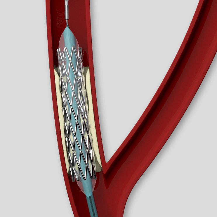
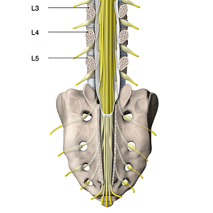
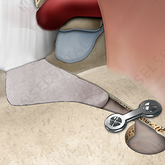
Visualisierungen für Patienten­informationen, Fach­publikationen, Lehrmittel und andere Formen der Wissenschafts­kommunikation

„Wissenschaftliche Illustrationen verdeutlichen, erklären und machen Unsichtbares sichtbar“

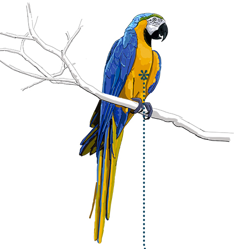
Je nach Bedürfnis des Kunden und Zweck der Abbildung wird eine entsprechende Bildsprache gewählt: von linear–schematisch bis plastisch–naturalistisch. Die Umsetzung erfolgt digital als klassische Illustration, 3D Visualisierung oder eine Kombination davon.

Ich freue mich über Ihre Anfrage:

anja.giger@bilderkram.ch

Beispielbilder im Portfolio ansehen

Blog & News

September 4, 2023

Published by Journal of Neurosurgery
American Association of Neurological Surgeons

Decompressive Craniotomy

Die Abbildung für Prof. Dr. med. Philippe Schucht hat es aufs Titelblatt des „Journals of Neurosurgery“ geschafft.

„JNS Vol 132 Issue 2“

Im Portfolio ansehen

Januar 9, 2023

The ‚worm‘ in our brain

In der Publikation „The ‚worm‘ in our brain. An anatomical, historical, and philological study on the vermis cerebelli“ – erschienen in „Journal of the History of the Neurosciences“ – beschreibt Klaus F. Steinsiepe die wandelnde Bedeutung und Interpretation des Vermis cerebelli seit der Zeit von Galen bis heute.

Ich gratuliere dem Autor zu dieser Publikation und freue mich, dass ich die Illustrationen zu diesem Artikel erstellen durfte.

„The cell doctrine—the theory of ventricular localization of the mental faculties—includes Galen’s idea of a locking or valve mechanism
between the middle and the rear ventricle (…) This article tells the story of the “worm’s” transformation from Galen to Vesalius and
beyond (…)“
Journal of the History of the Neurosciences 2023

Im Portfolio ansehen

September 2, 2022

Leonardo da Vinci, the Ventricles of the Brain, and the Foramen of Monro

In der Publikation „Leonardo da Vinci, the Ventricles of the Brain, and the Foramen of Monro“ , erschienen in „Nuncius – Journal of the Material and Visual History of Science“ – präsentieren Klaus F. Steinsiepe und Markus Hauser unter anderem mit grafischen Mitteln eine These, wie Leonardo da Vinci zu seiner Darstellung der Seitenventrikel gelangte.

Ich gratuliere den Autoren zu diesem spannenden Artikel und bedanke mich für die tolle Zusammenarbeit!

„Leonardo da Vinci’s neuroanatomical studies include a cast of the ventricles of an ox with melted wax. By depicting the result of this ingenious approach, he provided the first graphic representation of the connection between the cerebral ventricles.(…)“
Nuncius 2022

Im Portfolio ansehen

August 30, 2019

Craniotomy atlas

Zusammen mit Alain Blank durfte ich die Illustrationen für den „Craniotomy Atlas“ von Prof. Dr. med. Andreas Raabe erstellen. Nach mehrjähriger Arbeit ist das Buch am 10. Juli 2019 im Thiemeverlag erschienen.

„.. More than 600 high-quality operative photographs and brilliant illustrations support the step-by-step descriptions, with all the precision and attention to detail that neurosurgeons have come to expect from the editor Professor Raabe, and the associate editors Professors Meyer, Schaller, Vajkoczy, and Winkler…“ (Thieme)

Auswahlbilder ansehen

November 10, 2017

Brain

Abschlussarbeit Bildungsgang 3D Visualisierung & Animation | 2017

Extraktion eines groben Hirnmodelles aus einem MRI Bildsatz
Modellierung, Retopo, Texturierung und Animation mithilfe verschiedener 3D- und Animationsprogrammen

Ansehen